HER-2 Antibody Reagent for Immunohistochemistry
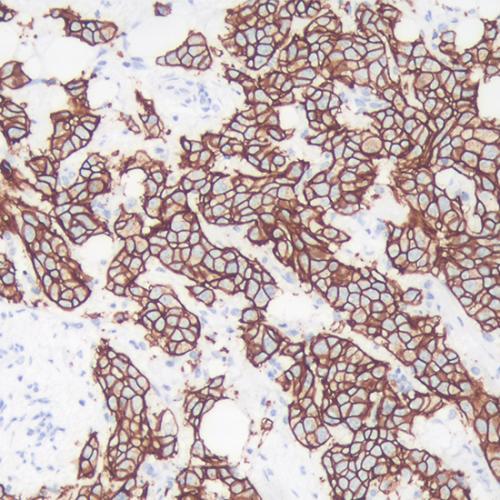
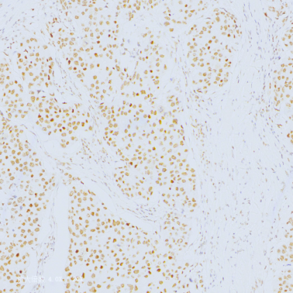
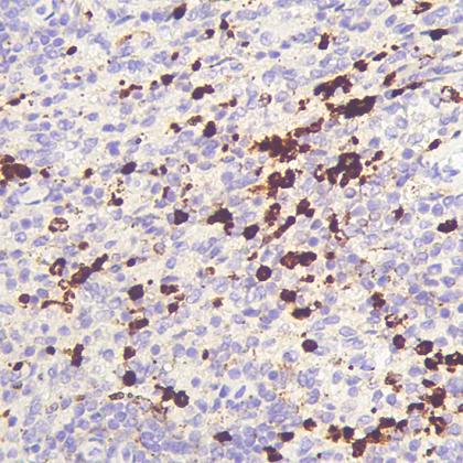
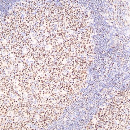
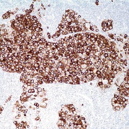
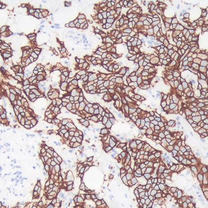
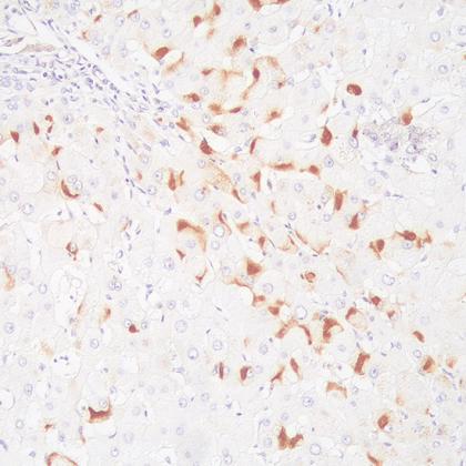
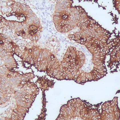
【Product name】 IHC Antibody -- HER-2 【Packing specification】 Code Clone Specifications AR3371 SP3 Concentrated:0.2ml RTU:1ml, 3ml, 6ml AM3071 e2-4001+3B5 AP3072 polyclonal 【Intended use】 For Research use only. HER-2 Antibody reagent is intended for use to qualitatively identify HER-2 by microscopy in sections of FFPE tissue using immunohistochemical detection system. 【Principle】 The HER-2 proto-oncogene is a transmembrane receptor tyrosine kinase that is clinically indicated in a number of carcinomas. Overexpression of the c-erbB-2 protein has been associated with ductal breast cancer, as well as pulmonary and gastric adenocarcinomas. A correlation between HER-2 and p53 has also been documented, as overexpression of both proteins has been associated with early invasion and metastasis in bladder cancer. Add the primary antibody to bind the antigen on tissue sections, and then use HRP labeled secondary antibody binding primary antibody to form the secondary antibody-primary antibody-antigen complex. When DAB chromogenic solution is added, HRP reacts with enzyme substrate to produce brown insoluble reaction product, which indirectly indicating the existence of antigen. 【Main components】 Immunoglobulin, antibody diluent 【Storage】 Store at 2~8℃ for 18 months. 【Sample requirements】 FFPE tissues are usually cut into sections as thin as 3~5μm with a microtome. These sections are then mounted onto glass slides that are coated with a tissue adhesive. 【Protocol】 1. Sample preparation:Deparaffinize the slides in xylene Ⅰ, Ⅱ, Ⅲ for 5 minutes;Transfer the slides once through 100%, 100%, 95%, 75% alcohols for 2 minutes respectively. Rinse slides with deionized water for 30 seconds. 2. Blocking:Block endogenous peroxidase activity by incubating sections in 3% H2O2 solution at room temperature for 5 minutes to block endogenous peroxidase activity. Rinse the slides with deionized water for 30 seconds. 3. Antigen retrieval:Heat the EDTA Antigen retrieval buffer to 100℃. Then place the slides in the boiled buffer and continue to heat for 15~20 min. Naturally cool down for 30 minutes. Rinse the sample with wash buffer. 4. Primary antibody incubation: Drain the slides. Add primary antibody to tissue, incubate at room temperature for 30 minutes. (use antibody diluent or PBS as control). Wash the slides in PBST for 2 times, 5 minutes for each time. If the Primary antibody is concentrated, please dilute it to RTU(ready to use) according to the information on packing. 5. Secondary antibody: Drain the slides. Add secondary antibody to tissue and incubate at room temperature for 20 minutes. Wash the slides in PBST for 2 times, 5 minutes for each time. 6. DAB:Drain the slides. Add DAB to the tissue and incubate at room temperature for 5 min. Rinse slides with deionized water. 7. Hematoxylin staining:Drain the slides. Add Hematoxylin to the tissue and incubate at room temperature for 5 minutes. Rinse slides with water. Use the acid solution for differentiation. Rinse slides with water. 8. Dehydrate:Dehydrate the slides in 75%, 95%, 100% alcohols for 2 minutes. Dry the slides. Cover stained tissue with a coverslip using mounting medium. 【Positive localization】 1. Positive localization: membrane. 2. Positive control:breast carcinoma. 【Precautions】 1. Please read the instruction carefully and become familiar with all components of the kit prior to use, Strictly follow the instruction during operation. 2. DO NOT use the kit or any kit component after their. 3. Only trained professionals can use this kit. Please wear suitable lab coat and disposable gloves while handling the reagents. 4. Avoid contact of skin, eyes and mucous membranes with the chemicals. 5. DO NOT pipet by mouth. 6. Unused reagents, used kit and waste must be disposed according to local regulations. 【Manufacturer】 Company Name: Xiamen Talent Biomedical Technology Co.,Ltd Address: The 3rd and 4th Floors, Building B10, No. 2068 Wengjiao West Road, BioMedical Park, Haicang District, Xiamen City, 361000 China Tel: +86 592 6315755 E-mail: talent1@talentbiomedical.com Website: www.talentbiomedical.com 【Symbols】